1 05 Graphic Display Of Electrocardiogram C Cardiac Rhythm

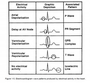
1 05 Graphic Display Of Electrocardiogram C Cardiac Rhythm C. electrical impulses of electrocardiogram waves. each part of the cardiac cycle produces a different electrical impulse. the electrical flow of the heart starts with the sa node (right atrium) and continues to the purkinje fibers (ventricles). electrodes transmit impulses to a recording pen which graphs the impulses in a series of up and down waves called deflection waves. 1 04. innervation; 1 05. graphic display of electrocardiogram (a) 1 05. graphic display of electrocardiogram (b) 1 05. graphic display of electrocardiogram (c) 1 05. graphic display of electrocardiogram (d) 1 05. graphic display of electrocardiogram (e) section iii. rhythms heart blocks. 1 06. analysis of ekg rhythm strips; 1 07. cardiac.

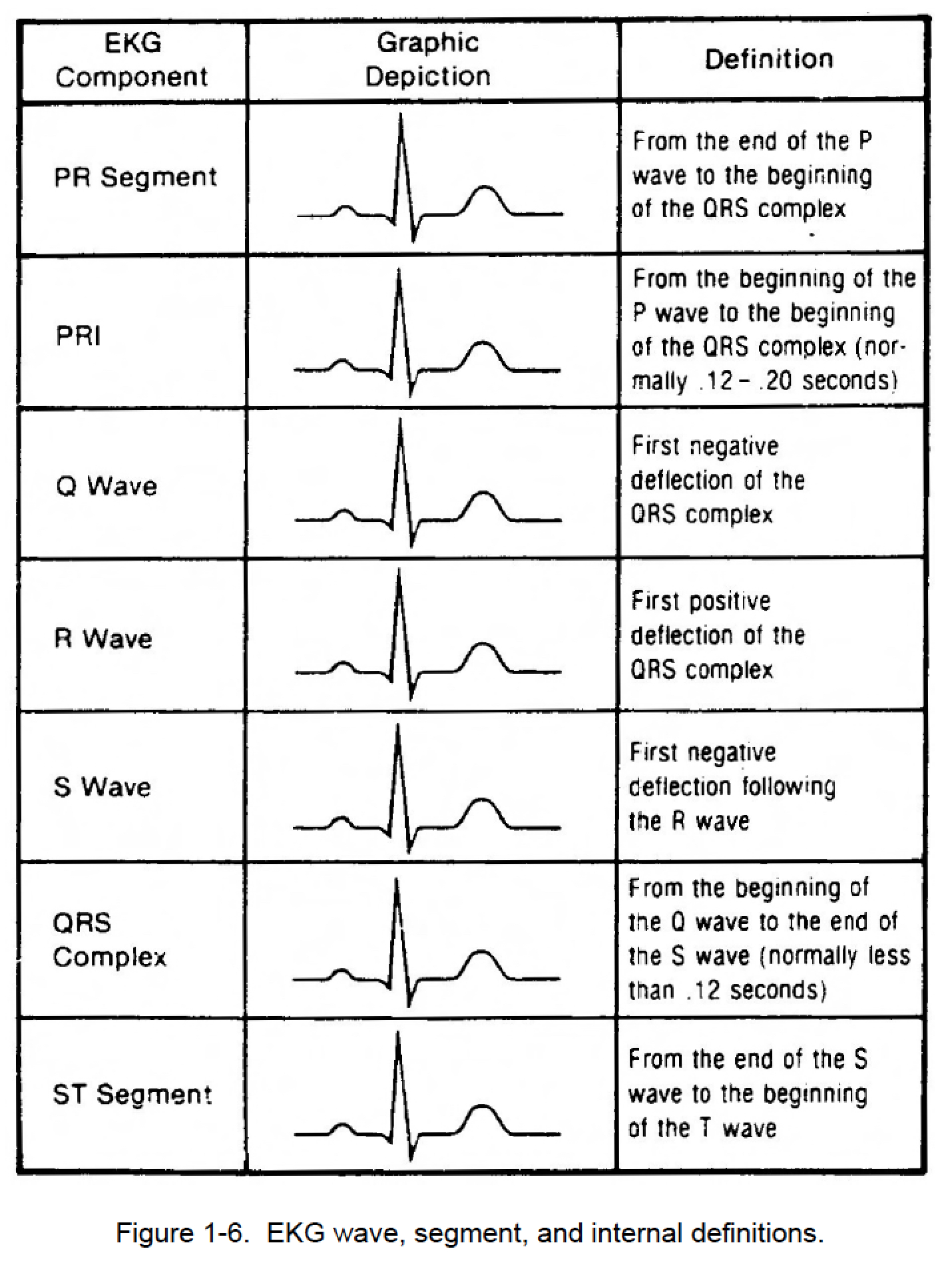
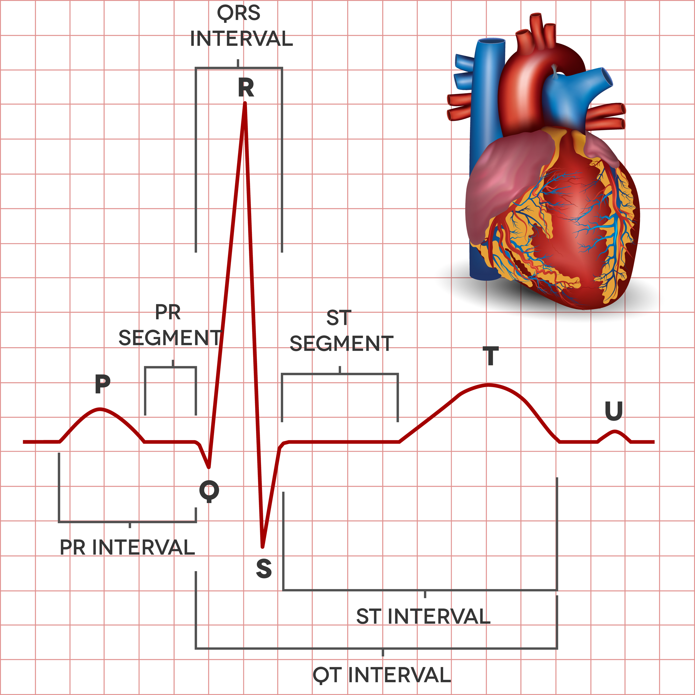
1 05 Graphic Display Of Electrocardiogram D Cardiac Rhythm A medical condition that disrupts this electrical impulse will display itself as a p r interval that is longer than 0.2 seconds due to the increased time it takes to travel the conducting tissues. the normal p r interval is between 0.12 and 0.20 seconds. schematic diagram of normal sinus rhythm for a human heart as seen on ecg (2) q wave. the q. A systematic approach to ecg interpretation: an efficient and safe method. the ecg must always be interpreted systematically. failure to perform a systematic interpretation of the ecg may be detrimental. the interpretation algorithm presented below is easy to follow and it can be carried out by anyone. the reader will gradually notice that ecg. Ecg rhythm evaluation. Electrocardiography (ecg) is a quick, simple, painless medical test that measures the heart’s electrical impulses. during an ecg, the heart's electrical impulses are measured, amplified, and recorded. this record, the electrocardiogram (also known as an ecg), provides information about the. part of the heart that triggers each heartbeat (the.

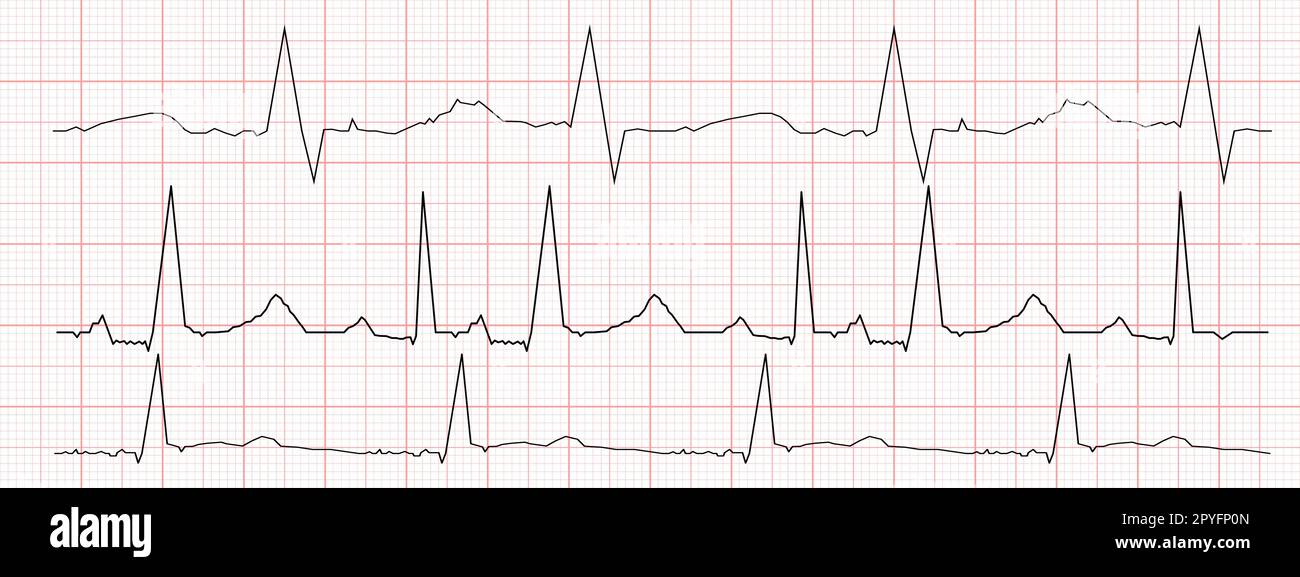
Set Of Black Heartbeat Diagrams On Red Graph Paper Ecg Ecg rhythm evaluation. Electrocardiography (ecg) is a quick, simple, painless medical test that measures the heart’s electrical impulses. during an ecg, the heart's electrical impulses are measured, amplified, and recorded. this record, the electrocardiogram (also known as an ecg), provides information about the. part of the heart that triggers each heartbeat (the. Frequent analysis of electrocardiogram (ecg) rate and rhythm provides for early identification and treatment of alterations in cardiac rhythm, as well as abnormal conditions in other body systems. this chapter presents a review of basic cardiac electrophysiology and information essential to the identification and treatment of common cardiac dysrhythmias. Dynamic electrocardiographic (ecg) monitoring is a standard of practice when providing general anesthesia, but opinions are mixed regarding its use during moderate (conscious) and deep sedation. the american dental society of anesthesiology included pulse oximetry for patient monitoring in its guidelines published in 1991. 1 the guidelines at.

The Electrocardiogram Explained What Is An Ecg Frequent analysis of electrocardiogram (ecg) rate and rhythm provides for early identification and treatment of alterations in cardiac rhythm, as well as abnormal conditions in other body systems. this chapter presents a review of basic cardiac electrophysiology and information essential to the identification and treatment of common cardiac dysrhythmias. Dynamic electrocardiographic (ecg) monitoring is a standard of practice when providing general anesthesia, but opinions are mixed regarding its use during moderate (conscious) and deep sedation. the american dental society of anesthesiology included pulse oximetry for patient monitoring in its guidelines published in 1991. 1 the guidelines at.

Ecg Patterns And Explanation

Comments are closed.