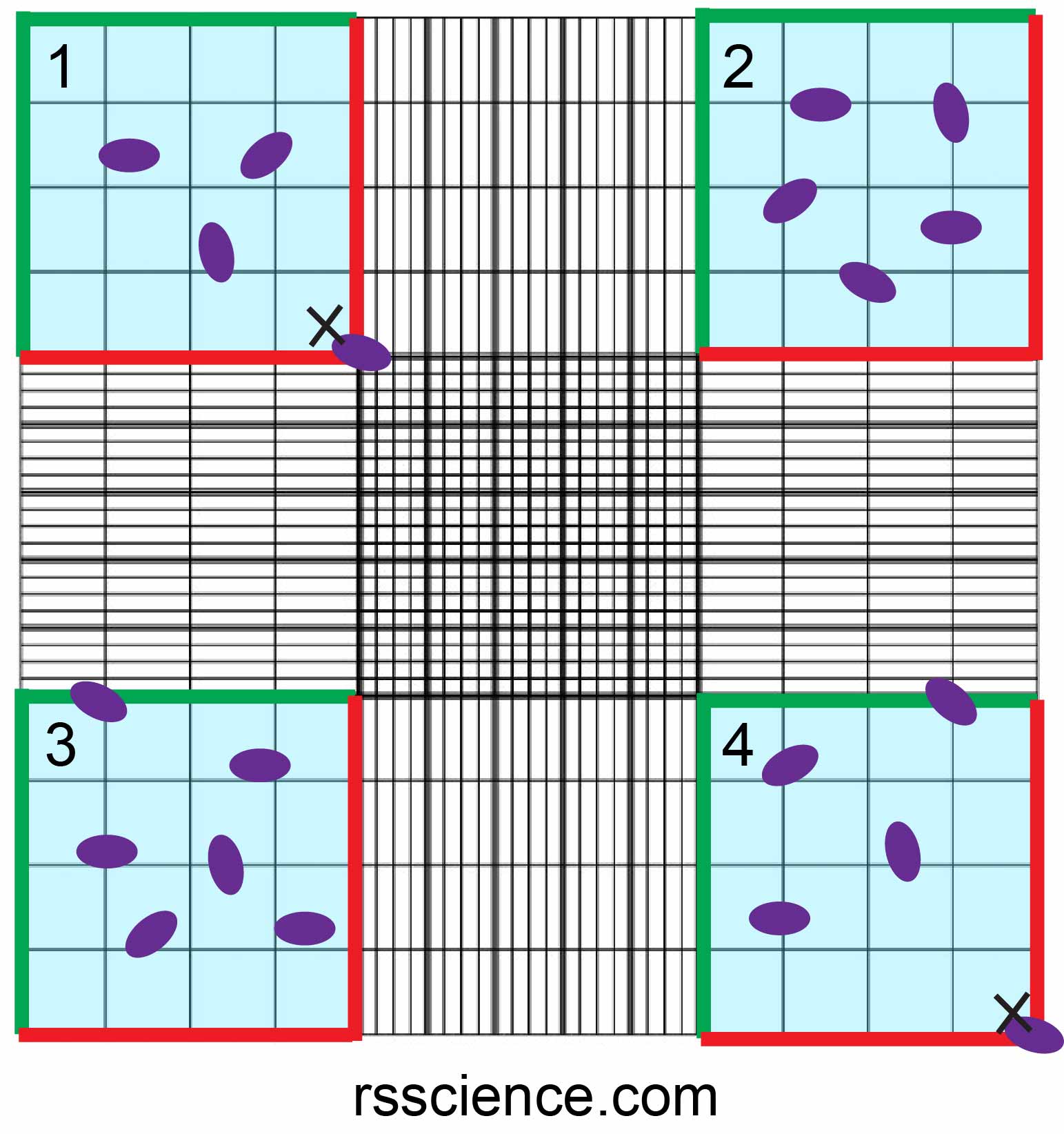
1 Haemocytometer The Blue Areas Indicate The Counting Areas Of The

1 Haemocytometer The Blue Areas Indicate The Counting Areas Of The Total area: 1 mm²; depth of chamber: 0.1 mm; volume of each small square: 0.0001 mm³ (0.1 nl) number of squares on the grid: 16; area of each small square: 1 16 mm² (0.0625 mm²) has two counting areas separated by a central barrier; the central barrier is 0.1 mm high, preventing mixing of the sample and reducing evaporation. Wbc count. when wbcs are counted, the calculation is much easier. wbcs are counted in the 4 corner squares of the main grid. these squares have an area of 1 mm2 each. to get the wbc count, the number of cells in each square are counted, and their mean is then calculated. let the mean be ‘n’. the volume of each square is 1 x 0.1 = 0.1 mm3.

Haemocytometer Showing The Counting Area Blue For Sperm Count And Using a microscope, focus on the grid lines of the counting area with a 5 10x objective. count the cells in one set of 16 squares (1×1 mm square area; the blue area). you should set a counting rule. for example, count the cells on the top and left lines of a square (marked by the green line), but do not count the cells on the bottom and right. How to count cells with a hemocytometer. Download scientific diagram | 1: haemocytometer the blue areas indicate the counting areas of the chamber from publication: whole thesis | label free biosensors are therefore an attractive. Steps. 1. using a pipette, take 100 µl of trypan blue treated cell suspension and apply to the hemocytometer. if using a glass hemocytometer, very gently fill both chambers underneath the coverslip, allowing the cell suspension to be drawn out by capillary action. if using a disposable hemocytometer, pipette the cell suspension into the well.

Hemocytometer Counting Chamber Medical Laboratory Medical Laboratory Download scientific diagram | 1: haemocytometer the blue areas indicate the counting areas of the chamber from publication: whole thesis | label free biosensors are therefore an attractive. Steps. 1. using a pipette, take 100 µl of trypan blue treated cell suspension and apply to the hemocytometer. if using a glass hemocytometer, very gently fill both chambers underneath the coverslip, allowing the cell suspension to be drawn out by capillary action. if using a disposable hemocytometer, pipette the cell suspension into the well. Remember if a cell overlaps a line, count it as “in” if it overlaps the top or right hand line and “out” if it overlaps the bottom or left hand line (figure 3f). the area of the middle (figure 3a) and each corner square (figure 3b–e) is 1 mm x 1 mm = 1 mm 2. the depth of each square is 0.1 mm. Focus on the grid lines of the haemocytometer using the 10×objective of the microscope. focus on one set of 16 corner squares as indicated by the circle. using a hand tally counter, count the number of cells in this area of 16 squares. when counting, always count only live cells that look healthy (unstained by trypan blue).

How To Use A Hemocytometer To Count Cells Rs Science Remember if a cell overlaps a line, count it as “in” if it overlaps the top or right hand line and “out” if it overlaps the bottom or left hand line (figure 3f). the area of the middle (figure 3a) and each corner square (figure 3b–e) is 1 mm x 1 mm = 1 mm 2. the depth of each square is 0.1 mm. Focus on the grid lines of the haemocytometer using the 10×objective of the microscope. focus on one set of 16 corner squares as indicated by the circle. using a hand tally counter, count the number of cells in this area of 16 squares. when counting, always count only live cells that look healthy (unstained by trypan blue).

Comments are closed.