Cavernous Sinus Contents And Cavernous Sinus Syndrome

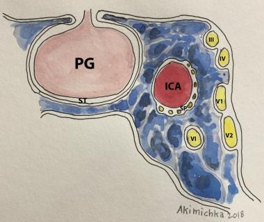
Cavernous Sinuses Usmle Strike Understanding the clinical management of cavernous sinus syndrome (ccs) requires an extensive understanding of its anatomy. it is a small but complex, and it contains several important structures. the cavernous sinus (cs) is not a venous plexus, but it is a true dural venous sinus.[1] it is bordered by the temporal bone of the skull and the sphenoid bone and lies lateral to the sella turcica. Cavernous sinus syndrome (css) is a condition caused by any pathology involving the cavernous sinus which may present as a combination of unilateral ophthalmoplegia (cranial nerve (cn) iii, iv, vi), autonomic dysfunction (horner syndrome) or sensory cn v 1 cn v 2 loss. anatomy. the venous drainage system of the head and face have a unique.

Imaging Spectrum Of Cavernous Sinus Lesions With Histopathologic The cavernous sinus. the cavernous sinus is a paired dural venous sinus located within the cranial cavity. it is divided by septa into small ‘caves’ – from which it gets its name. each cavernous sinus has a close anatomical relationship with several key structures in the head, and is arguably the most clinically important venous sinus. The cavernous sinus is part of the brain’s dural venous sinus and contains multiple neuro vasculatures. it is situated bilaterally to the sella turcica and extends from the superior orbital fissure anteriorly to the petrous part of the temporal bone posteriorly, and is about 1 cm wide and 2 cm long. the venous blood that flows to the cavernous sinus is from the superior and anterior. The cavernous sinus is a small but complex structure consisting of a venous plexus, the carotid artery, cranial nerves, and sympathetic fibers. broad categories of diseases involving the cavernous sinus can cause the so called cavernous sinus syndrome; these diseases include bacterial or fungal infections, noninfectious inflammation, vascular. Cavernous sinus syndrome (css) is a condition characterized by multiple cranial nerve palsies manifesting with ophthalmoplegia, ptosis, and facial sensory loss due to involvement of adjacent cranial nerves. tumors, trauma, and vascular, infectious, and noninfectious inflammatory disorders have all been described as causes.

Cavernous Sinus Syndromes Overview Clinical Presentation Diagnostic The cavernous sinus is a small but complex structure consisting of a venous plexus, the carotid artery, cranial nerves, and sympathetic fibers. broad categories of diseases involving the cavernous sinus can cause the so called cavernous sinus syndrome; these diseases include bacterial or fungal infections, noninfectious inflammation, vascular. Cavernous sinus syndrome (css) is a condition characterized by multiple cranial nerve palsies manifesting with ophthalmoplegia, ptosis, and facial sensory loss due to involvement of adjacent cranial nerves. tumors, trauma, and vascular, infectious, and noninfectious inflammatory disorders have all been described as causes. The cavernous sinus (cs) is not a venous plexus, but it is a true dural venous sinus. it is bordered by the temporal bone of the skull and the sphenoid bone and lies lateral to the sella turcica. the inferior and lateral walls and the roof of cs are extensions of the dura mater. there may or may not be a thin layer of collagen at the medial wall. Cavernous sinus thrombosis, a serious complication of paranasal sinusitis that most commonly results from the anterograde spread of infection involving the mid third of the face (e.g., orbit, mouth, paranasal sinuses), may be difficult to distinguish from simple orbital cellulitis.

Comments are closed.