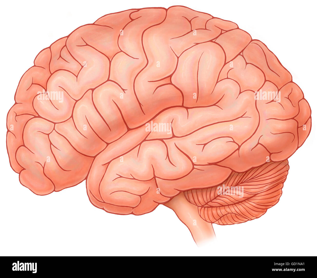
Im Gen De Un Cerebro Normal Download Scientific Diagram

Imгўgen De Un Cerebro Normal Download Scientific Diagram Download scientific diagram | imágen de un cerebro normal from publication: análisis sobre el reconocimiento automático de imágenes cerebrales | | researchgate, the professional network for. Download scientific diagram | imágenes spect simuladas de un cerebro normal (a), con lesiones compatibles con ea (b) y dm (c) a distintos tiempos de inyectado el rf (10 y 45 minutos).

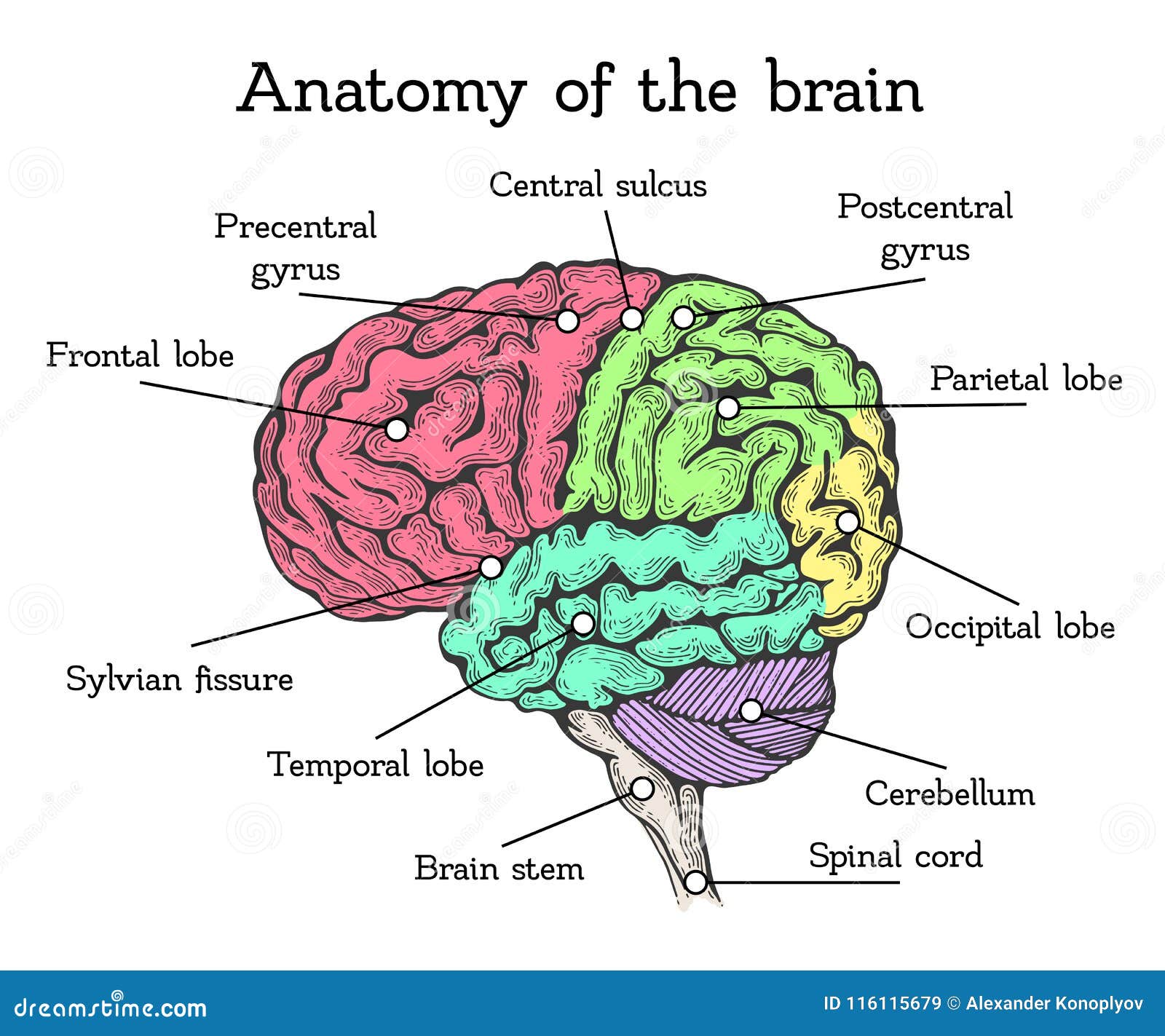
Normal Brain Cell Una anatomía topográfica del encéfalo para visualizar los diferentes niveles (encéfalo, diencéfalo, mesencéfalo, metencéfalo, protuberancia y cerebelo, rombencéfalo y prosencéfalo) junto con un diagrama de los diversos lóbulos cerebrales (lóbulo frontal, occipital, parietal, temporal, límbico e insular). Download scientific diagram | anatomía básica del cerebro. from publication: análisis de la difusión y curtosis aparentes en imágenes de resonancia magnética | el procesamiento digital de. 3d brain. for educators. log in. this interactive brain model is powered by the wellcome trust and developed by matt wimsatt and jack simpson; reviewed by john morrison, patrick hof, and edward lein. structure descriptions were written by levi gadye and alexis wnuk and jane roskams. like. subscribe. follow. Visores y reproductores. hhs divulgación de vulnerabilidad. national library of medicine 8600 rockville pike, bethesda, md 20894 u.s. department of health and human services national institutes of health. el cerebro es la porción más grande del sistema nervioso y se encuentra en la cavidad craneal.

Illustration Showing Anatomy Of A Normal Brain In A Superior Top View 3d brain. for educators. log in. this interactive brain model is powered by the wellcome trust and developed by matt wimsatt and jack simpson; reviewed by john morrison, patrick hof, and edward lein. structure descriptions were written by levi gadye and alexis wnuk and jane roskams. like. subscribe. follow. Visores y reproductores. hhs divulgación de vulnerabilidad. national library of medicine 8600 rockville pike, bethesda, md 20894 u.s. department of health and human services national institutes of health. el cerebro es la porción más grande del sistema nervioso y se encuentra en la cavidad craneal. 1.0 the nature and process of science. 2.0 introduction to human biology. 3.0 chemistry of life. 4.0 cells. 5.0 genetics. 6.0 biological evolution. 7.0 human evolution. 8.0 human variation. 9.0 introduction to the human body. Irm atlas axial del cerebro. atlas en línea gratuito con una serie completa de t1, t1, t2, t2 *, flair, imágenes axiales potenciadas por contraste de un cerebro humano normal. desplácese por las imágenes detalladas con etiquetas que utilizan nuestra interfaz interactiva. perfecto para los médicos, radiólogos y residentes que leen estudios de irm cerebral.

Diagrama De Anatomia Cerebral Con Secciones De Diferentes Colores Y Images 1.0 the nature and process of science. 2.0 introduction to human biology. 3.0 chemistry of life. 4.0 cells. 5.0 genetics. 6.0 biological evolution. 7.0 human evolution. 8.0 human variation. 9.0 introduction to the human body. Irm atlas axial del cerebro. atlas en línea gratuito con una serie completa de t1, t1, t2, t2 *, flair, imágenes axiales potenciadas por contraste de un cerebro humano normal. desplácese por las imágenes detalladas con etiquetas que utilizan nuestra interfaz interactiva. perfecto para los médicos, radiólogos y residentes que leen estudios de irm cerebral.

Comments are closed.