Imaging Of The Pelvis

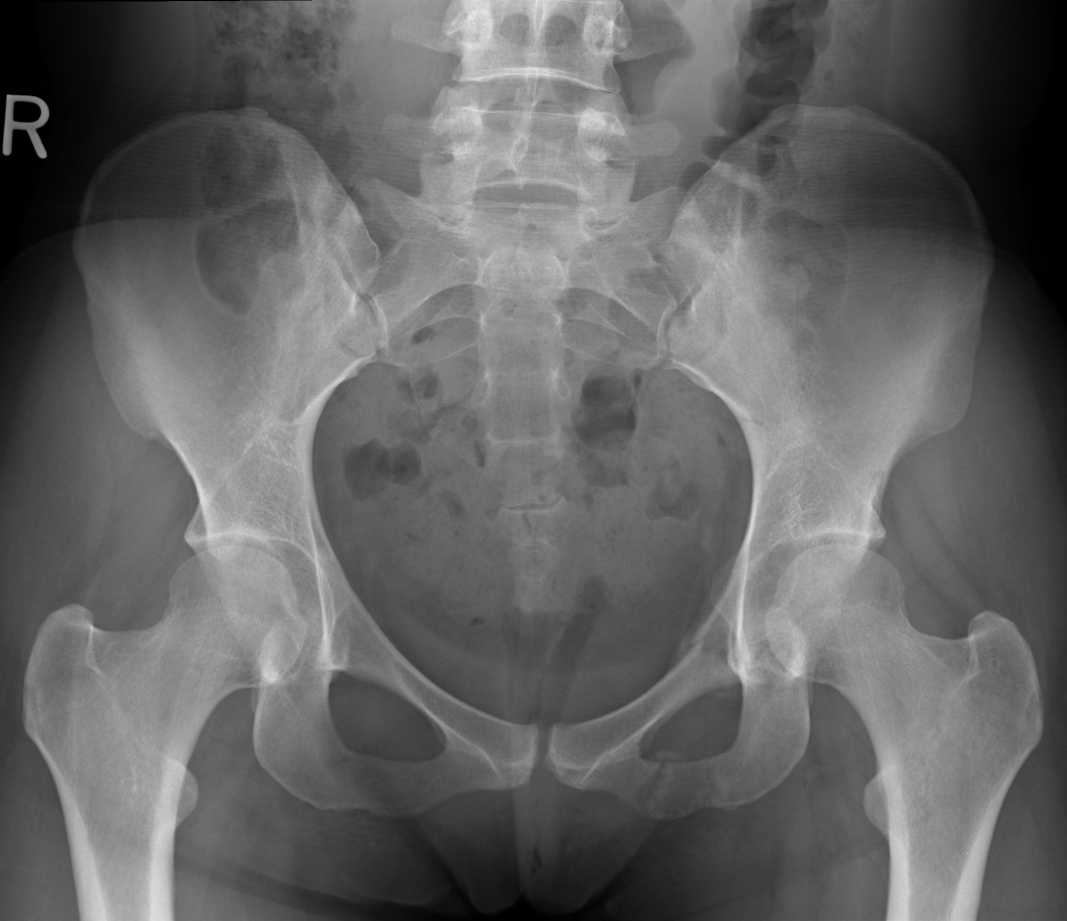
X Ray Of Normal Pelvis Female Eccles Health Sciences Library J Pelvic x ray and computed tomography techniques . the routine initial view of the pelvis is the anterior–posterior (ap) x ray ( figure 13 1 ).this image is obtained with the patient supine and the x ray beam oriented 90 degrees to the patient’s long axis, passing through the patient from anterior to posterior. What you need to know about pelvic mri.

Pelvic Anatomy Mri Human Anatomy Imaging the female pelvis: when should mri be considered?. Mri based synthetic ct can create hu maps and allows for automated segmentation of pelvic bones. the current and cutting edge mr techniques for bone imaging are complementary in the characterization of a variety of musculoskeletal disorders. keywords: pelvic bones, magnetic resonance imaging (mri), gradient echo mri, susceptibility weighted mri. T1 coronal view. magnetic resonance imaging or mri of the female pelvis offers a unique display of the pelvic anatomy, including a woman’s ovaries, uterus, and fallopian tubes. mri is a valuable technique in diagnosing or staging anomalies or conditions in the female pelvic region. unlike sonography or computed tomography (ct), mri offers. Magnetic resonance imaging (mri) offers infinite gray scale resolution allowing for improved tissue characterization and multiplanar capabilities. this facilitates the diagnosis of a variety of benign and malignant conditions involving the female pelvis without the use of ionizing radiation. common indications for mri of the pelvis are pain.

Magnetic Resonance Imaging Of The Pelvis T2 Weighted Sagittal View T1 coronal view. magnetic resonance imaging or mri of the female pelvis offers a unique display of the pelvic anatomy, including a woman’s ovaries, uterus, and fallopian tubes. mri is a valuable technique in diagnosing or staging anomalies or conditions in the female pelvic region. unlike sonography or computed tomography (ct), mri offers. Magnetic resonance imaging (mri) offers infinite gray scale resolution allowing for improved tissue characterization and multiplanar capabilities. this facilitates the diagnosis of a variety of benign and malignant conditions involving the female pelvis without the use of ionizing radiation. common indications for mri of the pelvis are pain. Practical approach to mri of female pelvic masses | ajr. General protocol. mr imaging of the female pelvis can be performed at 1.5 or 3.0 t field strength, although 3.0 t results in higher spatial resolution, superior signal to noise ratio, and shorter acquisition time. a multichannel phased array surface coil allows for parallel imaging that increases spatial resolution and decreases imaging time.

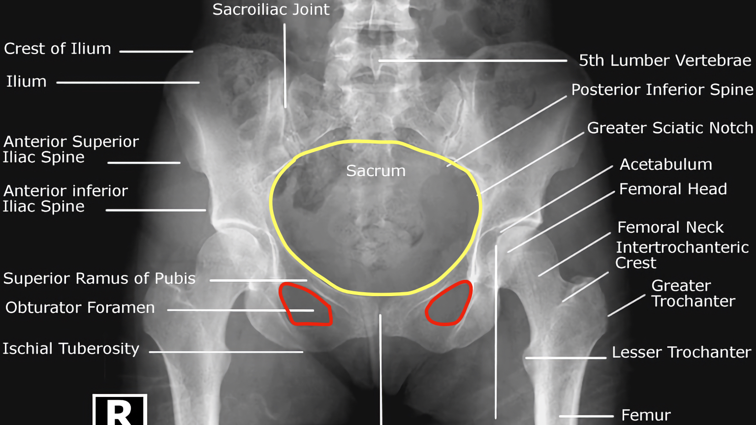
Hip X Ray Interpretation Osce Guide Geeky Medics Practical approach to mri of female pelvic masses | ajr. General protocol. mr imaging of the female pelvis can be performed at 1.5 or 3.0 t field strength, although 3.0 t results in higher spatial resolution, superior signal to noise ratio, and shorter acquisition time. a multichannel phased array surface coil allows for parallel imaging that increases spatial resolution and decreases imaging time.

Comments are closed.