Pathology Of Calcifying Aponeurotic Fibroma Rheumatoid Nodules

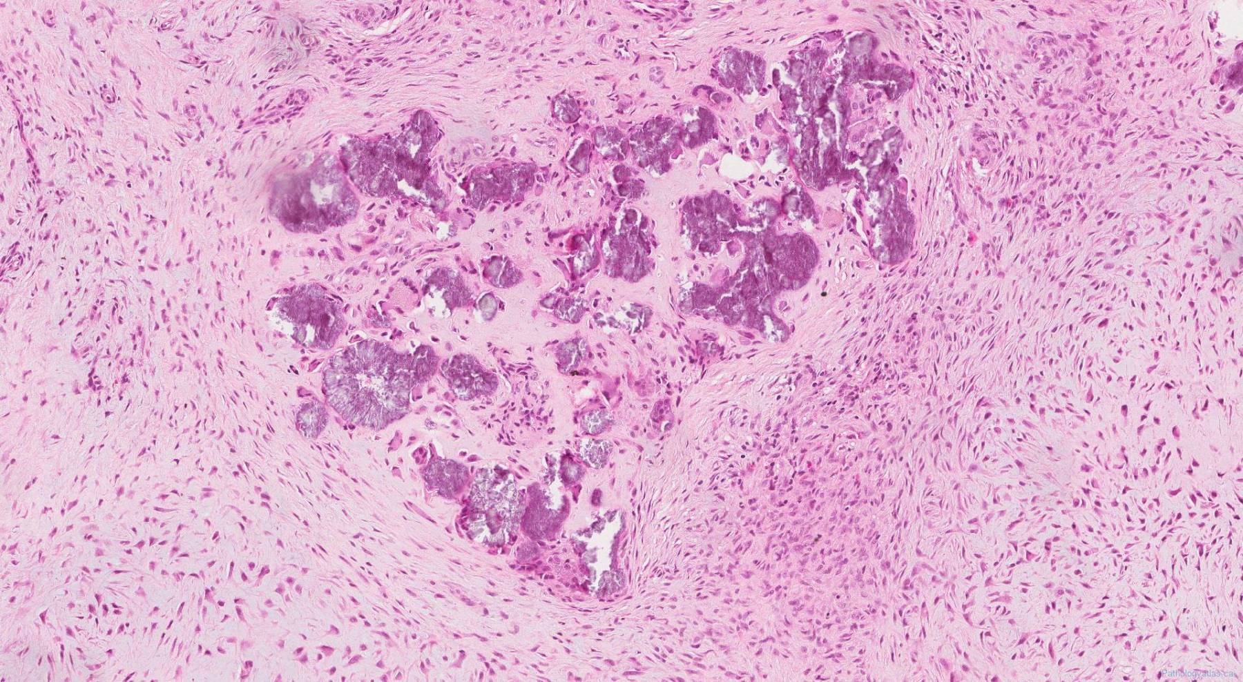
Pathology Outlines Calcifying Aponeurotic Fibroma In calcifying aponeurotic fibroma, sections show an irregularly shaped dense mass of fibrous tissue with foci of calcification (figures 1, 2). the fibrous tissue is quite cellular and appears to infiltrate the surrounding adipose tissue (figure 1). rarely extramedullary haematopoiesis may be seen. figure 1. Definition. calcifying aponeurotic broma (caf) is a rare. fi. tumor, usually occurring on the hands or feet, especially in the rst and second decades of life. fi. it is inltrative in nature, so has a relatively high. fi. local recurrence rate.

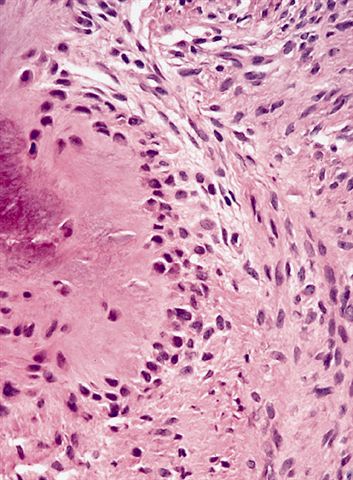
Pathology Outlines Calcifying Aponeurotic Fibroma Calcifying aponeurotic fibroma was first described and referred to as juvenile aponeurotic fibroma by keasbey in 1953 . it is a rare, benign, locally aggressive fibroblastic soft tissue tumor that typically occurs in the palm of the hand and in the sole of the feet in children and adolescents (1 3). the lesion has a tendency to infiltrate the. Abstract. we describe the first case of diagnosis of generalized calcifying aponeurotic fibroma in 52 year old man receiving long term therapy for seronegative rheumatoid arthritis with rheumatoid nodules. the prevalence of lesions (presence of multiple subcutaneous nodules in the aponeuroses and fascia of the head, neck, trunk, upper and lower. The nodules are well circumscribed by a proliferation of bland spindle cells and are composed of metaplastic cartilage with characteristic speckled irregular calcifications in a concentric fashion. this is the hallmark of calcifying aponeurotic fibroma (he, original magnification × 100). Calcifying aponeurotic fibroma was described as 'juvenile aponeurotic fibroma' by keasby in 1953. it is a rare, locally aggressive fibroblastic lesion located in the palms of the hands and soles of the feet in young children. these lesions have a recurrence rate of greater than 50% following surgical resection.

Pathology Of Calcifying Aponeurotic Fibroma Rheumatoid Nodules The nodules are well circumscribed by a proliferation of bland spindle cells and are composed of metaplastic cartilage with characteristic speckled irregular calcifications in a concentric fashion. this is the hallmark of calcifying aponeurotic fibroma (he, original magnification × 100). Calcifying aponeurotic fibroma was described as 'juvenile aponeurotic fibroma' by keasby in 1953. it is a rare, locally aggressive fibroblastic lesion located in the palms of the hands and soles of the feet in young children. these lesions have a recurrence rate of greater than 50% following surgical resection. Caf is a biphasic neoplasm composed of moderately cellular and infiltrative, fibromatosis like areas and nodules of calcification associated with more rounded epithelioid cells often radiating from the center of the calcifications. tumor cells lack significant atypia and show only rare mitotic figures. multinucleated osteoclast like giant cells. 'calcifying aponeurotic fibroma' published in 'encyclopedia of pathology' the microscopic findings vary little among caf cases. caf is most often biphasic lesion that displays a moderately cellular and infiltrative, fibromatosis like component and nodules of calcification accompanied by more rounded, epithelioid cells (fig. 1).

Calcifying Aponeurotic Fibroma Archives Atlas Of Pathology Caf is a biphasic neoplasm composed of moderately cellular and infiltrative, fibromatosis like areas and nodules of calcification associated with more rounded epithelioid cells often radiating from the center of the calcifications. tumor cells lack significant atypia and show only rare mitotic figures. multinucleated osteoclast like giant cells. 'calcifying aponeurotic fibroma' published in 'encyclopedia of pathology' the microscopic findings vary little among caf cases. caf is most often biphasic lesion that displays a moderately cellular and infiltrative, fibromatosis like component and nodules of calcification accompanied by more rounded, epithelioid cells (fig. 1).

Comments are closed.