Pathology Outlines Mucocele

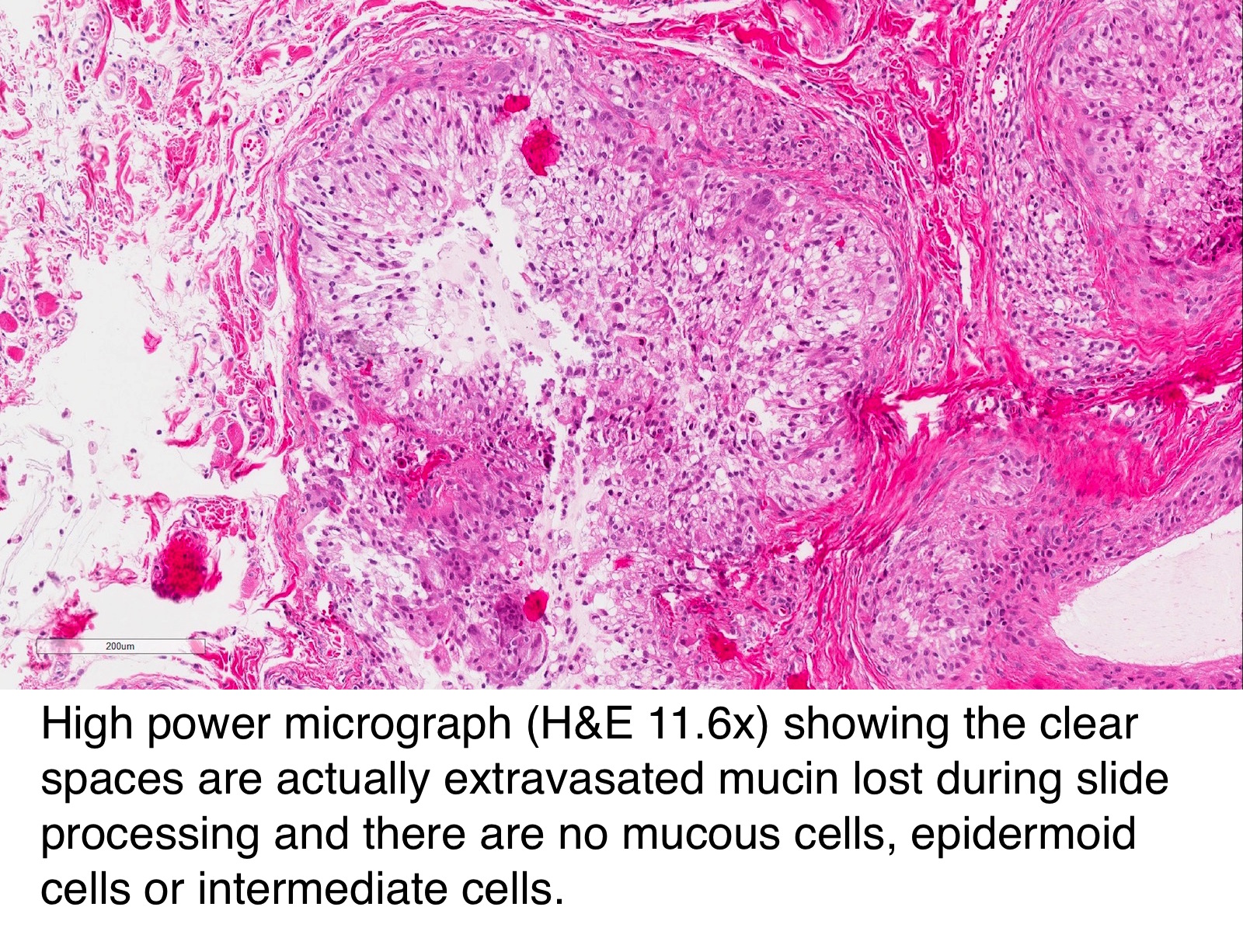
Pathology Outlines Mucocele Microscopic (histologic) description. microscopic features of mucocele depend on the underlying pathology. crypts may be decreased in the lamina propria but otherwise show no atypia (plos one 2017;12:e0179216) acellular mucin may dissect into the wall even in simple mucoceles (turk j gastroenterol 2020;31:649). Definition general. distended gallbladder containing clear and watery (hydrops) or mucoid secretions (mucocele), instead of bile. adult cases almost always due to impacted stones in ampulla or cystic duct. rarely due to regional tumors causing compression. pediatric cases associated with kawasaki syndrome or other inflammatory narrowing of.

Pathology Outlines Mucocele Oral cavity salivary duct cyst. complete excision along with the feeding minor salivary gland is curative (arch otolaryngol head neck surg 1987;113:51) iatrogenic intraoperative damage to neighboring salivary gland parenchyma may contribute to the development of postoperative mucocele (oral surg oral med oral pathol 1964;18:191). Salivary gland mucocele. salivary gland mucocele, also salivary mucocele and mucocele, is a benign lesion of the head and neck. ranula redirects here. a ranula is a mucocele that occurs in the floor mouth. [1]. Oral mucocele is the most common benign minor (accessory) salivary gland lesion, caused due to mechanical trauma to the excretory duct of the gland. clinically they are characterized by single or multiple, soft, fluctuant nodule, ranging from the normal color of the oral mucosa to deep blue. it affects at any age and is equally present in both. Mucoceles and ranulas are among the most common disorders of the salivary glands. mucocele, which is of minor salivary gland origin, arises when there is a disruption of the flow of its secretions. mucoceles are of two types: extravasation mucoceles and retention mucoceles. the former results mostly from trauma to the salivary duct, leading to the collection of secretions in the connective.

Pathology Outlines Mucocele Oral mucocele is the most common benign minor (accessory) salivary gland lesion, caused due to mechanical trauma to the excretory duct of the gland. clinically they are characterized by single or multiple, soft, fluctuant nodule, ranging from the normal color of the oral mucosa to deep blue. it affects at any age and is equally present in both. Mucoceles and ranulas are among the most common disorders of the salivary glands. mucocele, which is of minor salivary gland origin, arises when there is a disruption of the flow of its secretions. mucoceles are of two types: extravasation mucoceles and retention mucoceles. the former results mostly from trauma to the salivary duct, leading to the collection of secretions in the connective. Mucocele. mucocele is cavity that contains a mucous secretion. it can be either benign or malignant. [1] [2] it may refer to: salivary mucocele. Introduction. although a distended, mucus filled appendix is often called a mucocele, this term is ambiguous and best utilized to describe an imaging appearance rather than a pathologic entity. the underlying biology and behavior of appendiceal mucinous lesions are variable and can range from non neoplastic to neoplastic.

Comments are closed.