Quick 12 Lead Ekg Review Location Of Infarct And Which Artery Is Affected

Quick 12 Lead Ekg Review Location Of Infarct And Which Just a quick video on how on a method to use while looking at an ekg to determine which part of the heart and which coronoary artery is affected. Ecg localization of myocardial infarction ischemia and.

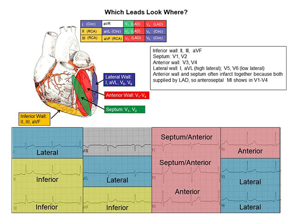
12 Lead Ecg Reference Chart вђ Cardiovascular Nursing Education Associates Precordial leads. v1 and v2 are on either side of sternum at 4th ics. v4 is midclavicular line, 5th ics. v3 is halfway between v2 and v4. v6 is at midaxillary line, 5th ics. v5 is halfway between v4 and v6, 5th ics. 12 lead ecg. Pathophysiology. occlusion of a coronary artery disrupts the blood supply to a region in the myocardium. ischemia ensues, the myocytes become rapidly dysfunctional. when ischemia persists, this can result in myocyte death. after 30 minutes of severe ischemia, the damage becomes irreversible. infarction patterns. Coronary arteries. rca – inferior lv ecg leads: il, lll, avf. l cx lateral lv ecg leads: i, avl, v5, v6. lad – anterior lv ecg leads: v1 – v4. the coronary arteries deliver oxygen rich blood to the muscle tissues of the heart. if the arteries become blocked, heart muscle will die resulting in a heart attack. ecg electrode placement. Cases include text, audio and assessment exercises. we provide online access to courses, drills and quizzes to complement to classroom instruction. click to learn more. learn ecg, blood pressure measurement, lung sounds, and heart sounds. our mostly free training includes drills, quizzes and reference guides. for all medical professionals.

Comments are closed.