The Ureters Anatomical Course Neurovascular Supply Teachmeanatomy

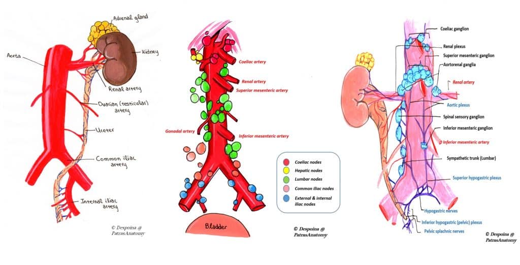
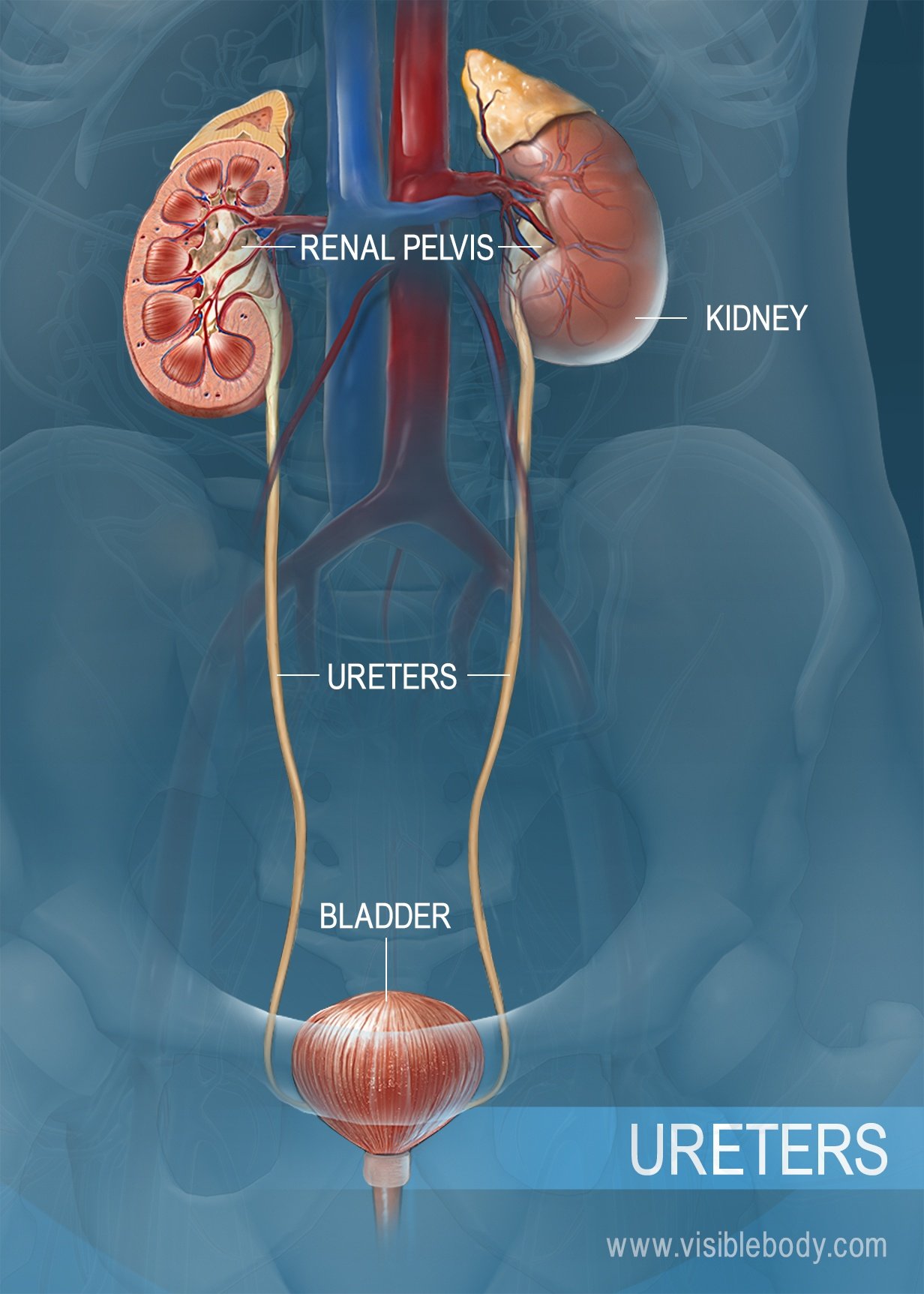
The Ureters Anatomical Course Neurovascular Supply Teachmeanatomy The ureters. the ureters are two thick tubes which act to transport urine from the kidney to the bladder. they are approximately 25cm long and are situated bilaterally, with each ureter draining one kidney. in this article, we shall look at the anatomy of the ureters – their anatomical course, neurovascular supply and clinical correlations. The urethra male female anatomical course.

The Ureters Anatomical Course Neurovascular Supply Teachmeanatomy Neurovascular supply. the rectum receives arterial supply through three main arteries: superior rectal artery – terminal continuation of the inferior mesenteric artery. middle rectal artery – branch of the internal iliac artery. inferior rectal artery – branch of the internal pudendal artery. venous drainage is via the corresponding. Ureters: anatomy, innervation, blood supply, histology. 🌟 in this educational masterpiece, we dissect the complete anatomy of the ureter, covering its course, constrictions, relations, blood supply, gender differ. The ureters are bilateral thin tubular structures with a 3 to 4 mm diameter that connect the kidneys to the urinary bladder (see image. posterior thoracolumbar surface anatomy). these muscular tubes transport urine from the renal pelvis to the bladder. the ureter's muscular layers are responsible for the peristaltic activity that moves urine from the kidneys to the bladder.

Structure And Function Of The Ureters 🌟 in this educational masterpiece, we dissect the complete anatomy of the ureter, covering its course, constrictions, relations, blood supply, gender differ. The ureters are bilateral thin tubular structures with a 3 to 4 mm diameter that connect the kidneys to the urinary bladder (see image. posterior thoracolumbar surface anatomy). these muscular tubes transport urine from the renal pelvis to the bladder. the ureter's muscular layers are responsible for the peristaltic activity that moves urine from the kidneys to the bladder. Ureter. the anatomy of the urinary system consist of: kidneys, ureters, bladder and urethra. urine is created in the renal tubules, and it is stored in the kidney’s renal pelvis. urine flows from the kidneys, passing through the ureters to the bladder. urine builds up in the bladder until it is ejected from the body through the urethra. Superior mesenteric arteries and veins, ovarian and testicular vessels, as well as the mesentery of the sigmoid colon and parietal peritoneum pass anteriorly to it. the length of the ureters of an adult is approximately 35 cm. the ureter consists of three parts: 1. abdominal part (pars abdominalis) abdominal part.

Comments are closed.