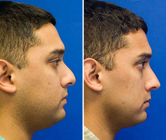
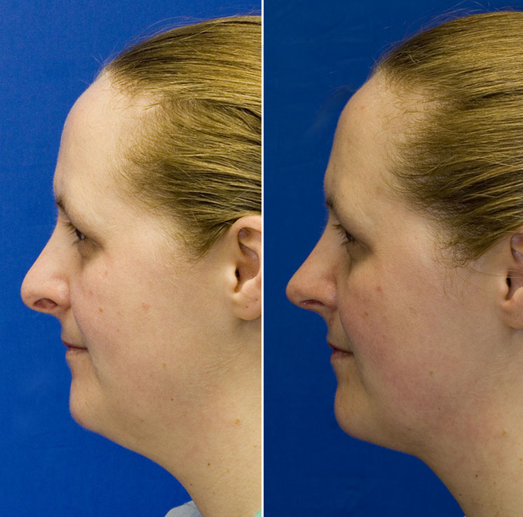
What Is A Pollybeak Deformity

Pollybeak Deformity Rhinoplasty In Seattle Rhinoplasty Surgeon Polly beak deformity in rhinoplasty. What is a pollybeak deformity? a pollybeak deformity occurs when the area above the tip of the nose on the bridge (also called the supratip) is the highest part of the nose when seen from a profile view. the pollybeak term comes from how the nose looks like a parrot beak. causes of a pollybeak deformity. a pollybeak deformity can occur from a.

Pollybeak Deformity Rhinoplasty In Seattle Rhinoplasty Surgeon Pollybeak deformity refers to excess tissue over the supratip area (region over the bridge of the nose right before the nasal tip). this causes the nose to be beak shaped in appearance, hence the name used for this deformity. this usually occurs from prior rhinoplasty surgery. Pollybeak occurs when the swelling or tissue in the supratip pushes the nose downward into the beak like formation. this deformity is a serious imperfection that will not resolve on its own and require revision surgery to adjust the cartilage and bridge for a natural and balanced nose. A pollybeak deformity is when the supratip area of the nose – which is the nasal bridge area that comes right before the nasal tip – appears full from excess tissue. this fullness in the supratip region creates a nose that resembles a parrot’s beak, hence the name of this deformity. What is a pollybeak deformity? as the name suggests, a pollybeak deformity is when the nose resembles the shape of a bird beak when looked at from the profile view. the supratip – which is the area of the nose located between the nose tip and the saddle – is fuller and more prominent than the tip, causing this visual deformity.

Pollybeak Deformity Rhinoplasty In Seattle Rhinoplasty Surgeon A pollybeak deformity is when the supratip area of the nose – which is the nasal bridge area that comes right before the nasal tip – appears full from excess tissue. this fullness in the supratip region creates a nose that resembles a parrot’s beak, hence the name of this deformity. What is a pollybeak deformity? as the name suggests, a pollybeak deformity is when the nose resembles the shape of a bird beak when looked at from the profile view. the supratip – which is the area of the nose located between the nose tip and the saddle – is fuller and more prominent than the tip, causing this visual deformity. Polly beak deformity, also called supratip fullness, is a condition that occurs when there is an overgrowth of the scar, fat, or cartilage in the supratip area of the nasal bridge, causing a visible bump or fullness in that area. (1) this deformity exposes cartilage, which appears like a parrot's beak, and pushes the nasal tip downwards. If pollybeak deformity and dimpling deformity of the nasal tip is present, the supratip transposition flap is designed adjacent to the nasal tip depression (fig. 16). to minimize a standing cone deformity over the donor site, an ellipti cal flap, instead of a round one, is designed (fig. 16a).

Correction Of Pollybeak And Dimpling Deformities Of The Nasal Tip In Polly beak deformity, also called supratip fullness, is a condition that occurs when there is an overgrowth of the scar, fat, or cartilage in the supratip area of the nasal bridge, causing a visible bump or fullness in that area. (1) this deformity exposes cartilage, which appears like a parrot's beak, and pushes the nasal tip downwards. If pollybeak deformity and dimpling deformity of the nasal tip is present, the supratip transposition flap is designed adjacent to the nasal tip depression (fig. 16). to minimize a standing cone deformity over the donor site, an ellipti cal flap, instead of a round one, is designed (fig. 16a).

Comments are closed.